When someone experiences a traumatic event like a collision, a fall, or a workplace accident, every decision that follows matters. Accurate diagnosis, early detection, and clear documentation can influence not only a patient’s health outcomes but also the legal and financial support they receive. That’s why personal injury cases require more than standard imaging. They require precision diagnostics that reveal what basic scans often miss.
At SimonMed, we bring AI-enhanced, advanced imaging technologies to communities across the country, making world-class personal injury imaging more accessible, more accurate, and more affordable. Our goal is simple: Help people live healthier, longer lives, starting with the right diagnosis.
Navigate to:
- Understand traumatic brain injury
- Advanced brain imaging tools
- When to image after trauma
- The SimonMed advantage in spine imaging for personal injury
- Why personal injury attorneys and patients choose SimonMed
- A clearer diagnosis, a stronger case, a healthier future
Understanding traumatic brain injury: why standard MRIs miss so much
A traumatic brain injury (TBI) can be life-changing. But the reality is that standard MRI sequences often fail to show the subtle injuries that many people experience after trauma.
Traditional scans can “skip” tiny lesions when slices are too thick. Many clinics use 4–5 mm slices; SimonMed uses 1.2 mm thin-section imaging, capturing detail that would otherwise go unseen.
We pair this with:
- Multiplanar imaging, including additional sagittal FLAIR sequences
- 3.0 Tesla MRI magnets, the highest-quality imaging standard available
- Advanced protocols used in leading research centers
This combination ensures that if there’s an injury, we can see it, early and clearly.
Advanced brain imaging tools that matter in personal injury cases
To diagnose TBI with confidence, we use a suite of advanced MRI techniques designed to reveal different kinds of injury. Each method uncovers information that standard imaging simply cannot provide.
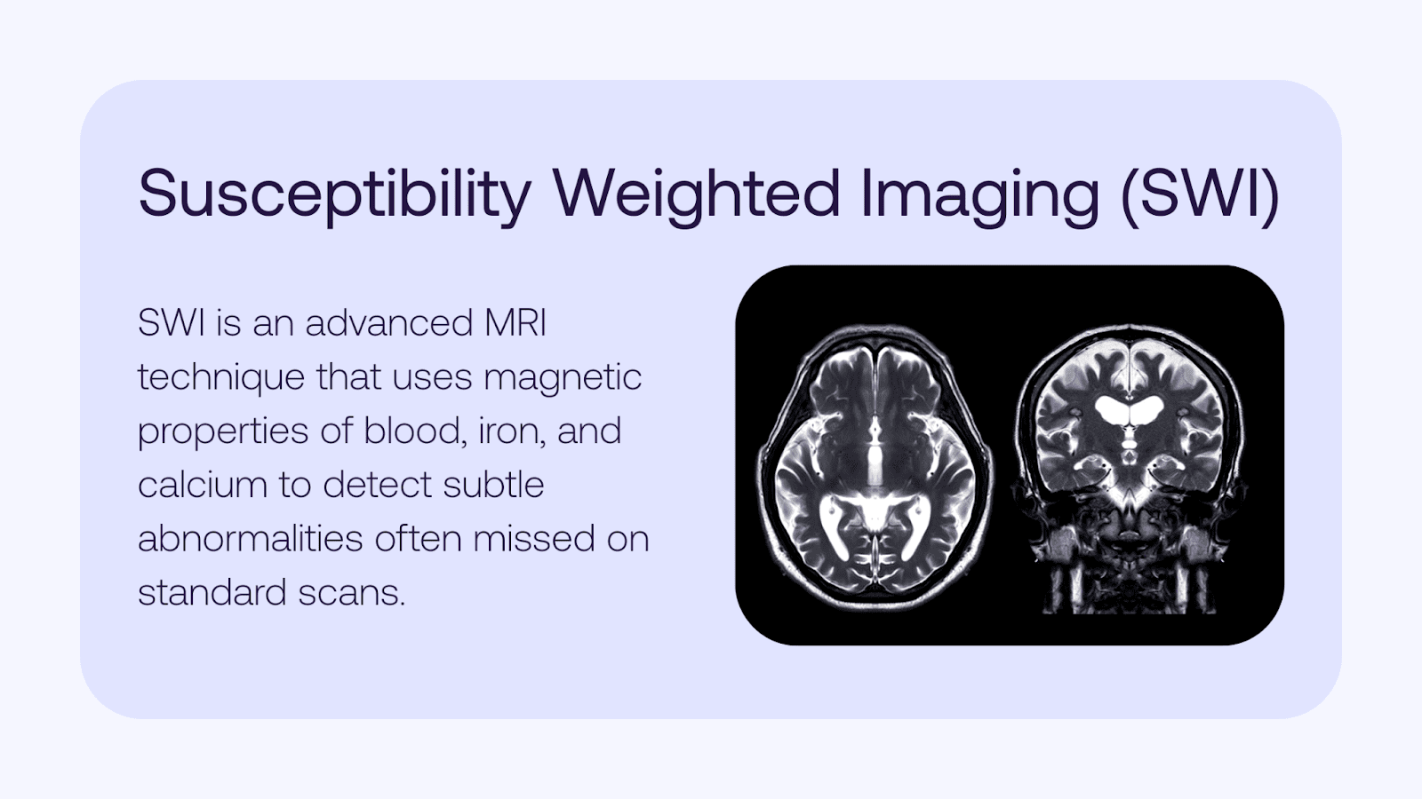
1. Susceptibility Weighted Imaging (SWI)
SWI is one of the most important tools in TBI detection. It identifies tiny amounts of hemorrhage, blood products, or calcium, even when other MRI sequences appear normal.
It excels at identifying:
- Diffuse axonal injury (DAI) microhemorrhages
- Shearing injuries from rotational trauma
- Hemorrhagic lesions that explain persistent symptoms
These microbleeds are often invisible on conventional MRI, which is why SWI is essential in personal injury work.
2. Diffusion Tensor Imaging (DTI)
DTI analyzes the brain’s white matter tracts that connect different parts of the brain.
This MRI technique detects:
- Microscopic damage
- Altered communication pathways
- Disrupted structural integrity of neural networks
Because traditional MRI is frequently insensitive to microscopic injury, DTI adds crucial diagnostic and prognostic value—especially in concussion or mild TBI cases.
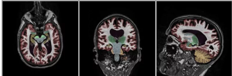
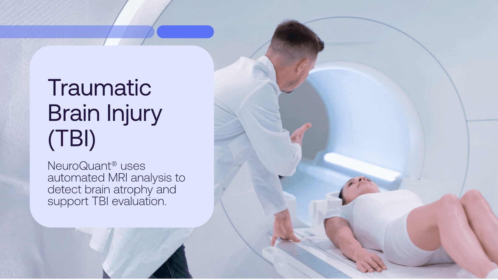
3. NeuroQuant® volumetrics
After trauma, the brain may swell, and then begin to atrophy. NeuroQuant is the first FDA-cleared, quantitative brain imaging analysis software that segments and measures volumes of brain structures and compares them to an age-matched database.
This helps radiologists:
- Detect early or subtle post-traumatic atrophy
- Track changes over time
- Quantify hippocampal, cortical, or deep gray matter loss
- Assess ventricle size as swelling resolves
Volumetric analysis provides objective, measurable data that strengthens both clinical care and legal clarity.
4. Perfusion Imaging
Perfusion MRI measures blood flow through the brain using a magnetic field. Abnormal perfusion patterns can indicate:
- Brain regions struggling after injury
- Chronic symptoms caused by poor oxygenation
- Vascular injury related to trauma
This adds another layer of insight and context beyond what structural imaging reveals.
5. Spectroscopy & fMRI
For select cases, we can evaluate:
- Cellular-level metabolic changes (MR spectroscopy)
- Functional activation patterns (fMRI)
These tools help identify subtle but meaningful disruptions in brain health and cognitive function.
When to image after trauma
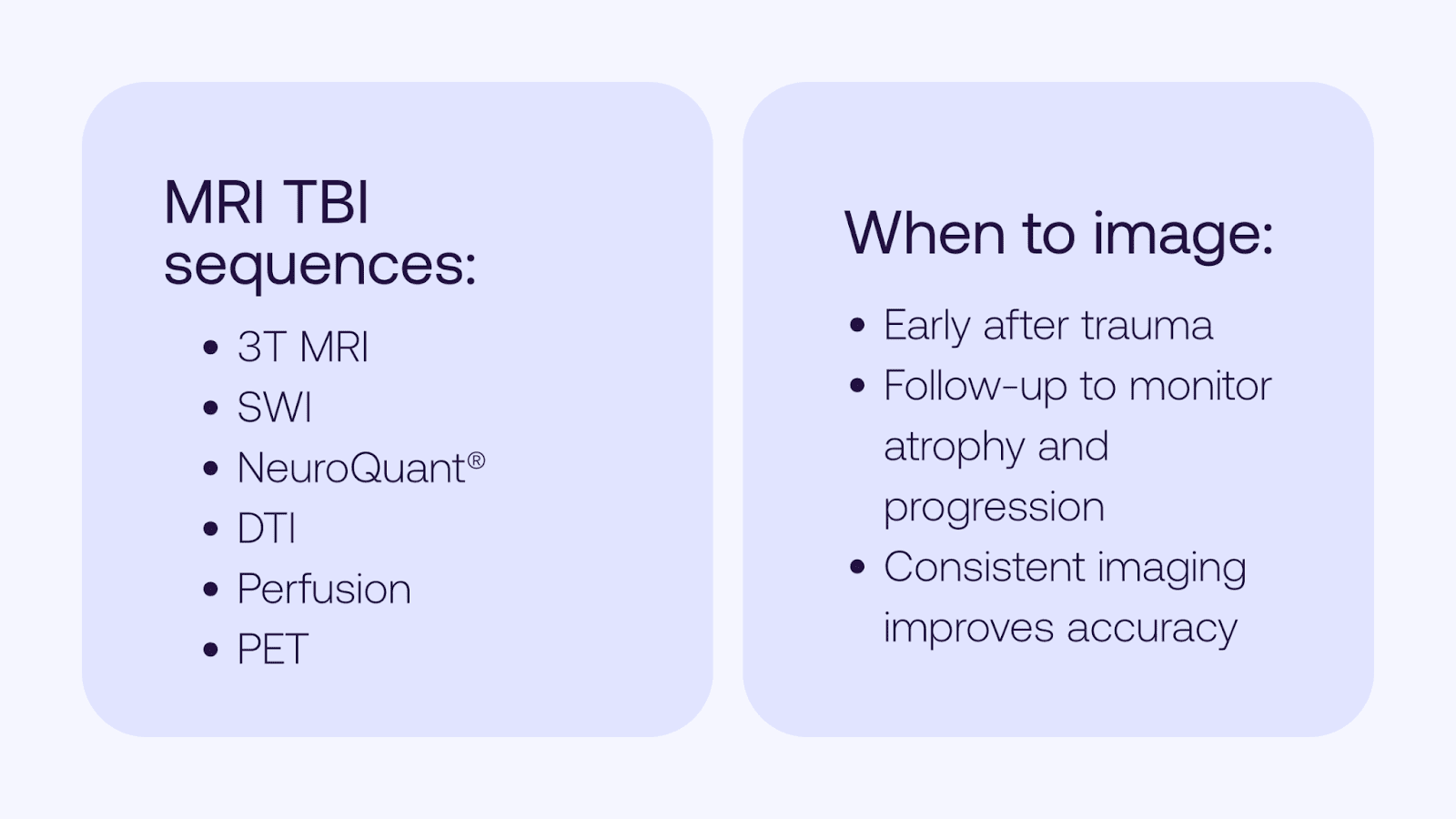
Timing matters. We follow evidence-based guidance that supports optimal diagnostic accuracy:
Immediately after trauma
- Detect hemorrhages
- Identify diffuse axonal injury
- Rule out urgent complications
Hemorrhages often resolve quickly, so early imaging captures findings that may fade within days.
1 Week – 3 months post-injury
The brain begins to show interval atrophy, peaking at 2–3 months and continuing for 6 months or more.
This is the ideal window to use:
- NeuroQuant
- DTI
- Perfusion imaging
Using the same scanner over time allows for precise, reliable tracking of changes.
The SimonMed advantage in spine imaging for personal injury
Brain injuries aren’t the only consideration in trauma. Spinal injuries can be just as disruptive, and just as subtle. We provide specialized spine imaging tailored for personal injury cases.
When to image the spine
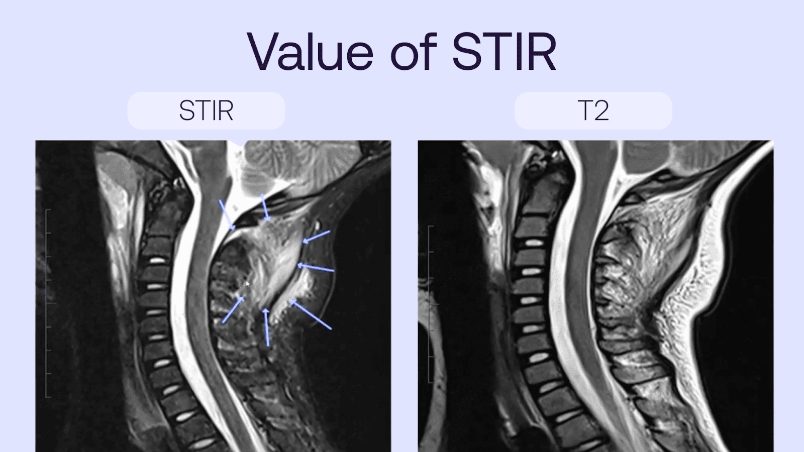
- Immediately when STIR imaging suggests ligament injury or edema
- For follow-up, to observe healing, edema resolution, or new osteophyte development
Dedicated spine sequences
We deliver some of the most advanced spine protocols available:
- 3.0 Tesla MRI
- STIR imaging for acute ligamentous injury
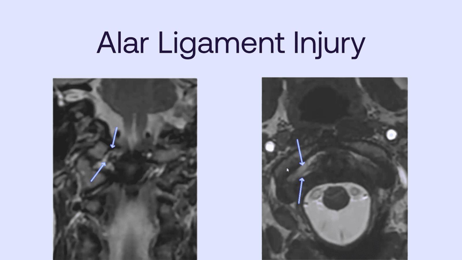
- Alar ligament imaging for craniocervical instability and whiplash
- Flexion-extension MRI to detect dynamic instability
- Spine DTI (in select cases)
Specialized spine X-rays
When needed, our team performs:
- Flexion-extension views
- Open-mouth odontoid imaging
- Side bending studies
- Dynamic video fluoroscopy
These tools reveal vertebral body motion, translation, and instability that standard films may miss.
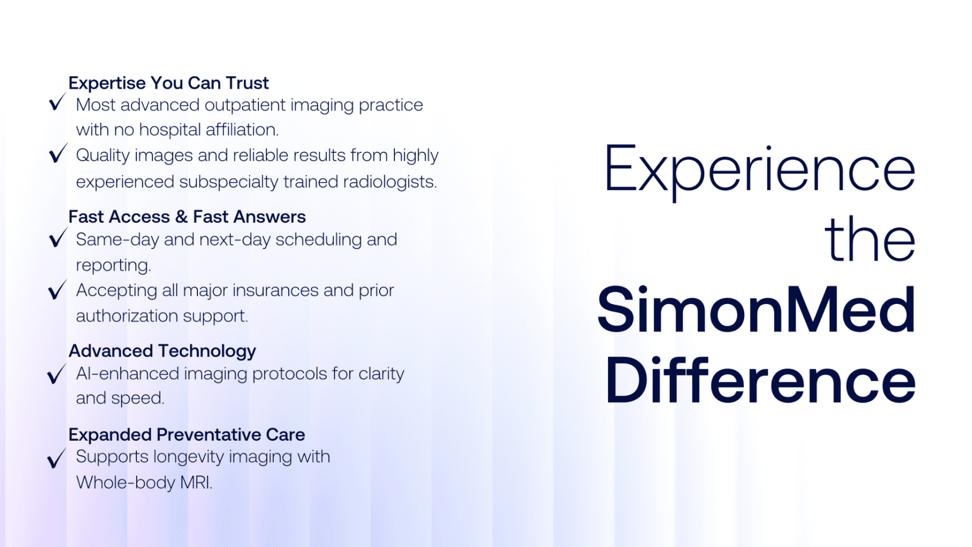
Why personal injury attorneys and patients choose SimonMed
We go far beyond the basics. Our goal is to provide a complete, precise picture—not a partial one.
The best standard sequences
- Thin slices (1.2 mm) so no lesions are “skipped”
- Extra sagittal FLAIR sequences
- 3T MRI magnets (like upgrading to the best camera lens)
- Top protocols for spine, MSK, and brain imaging
The best advanced sequences
Vendors often “lock” advanced sequences behind costly keys. Many imaging centers skip them to save time and money. SimonMed doesn’t.
Our advanced sequences include:
- SWI
- DTI with 3D
- NeuroQuant
- Perfusion
- Spectroscopy
- fMRI
These tools are essential for identifying the full spectrum of traumatic injury.
Expert neuroradiologists
We’re home to radiologists who specialize in head trauma, neuroimaging, and personal injury cases.
Our team provides:
- Expert witness support
- Research-backed trauma analysis
- Technologists with 10+ years’ experience in advanced imaging
Nationwide accessibility
As the largest outpatient imaging provider in the United States, we offer:
- Faster appointments
- Streamlined scheduling
- Support for paperwork and case management
- Consistent communication
We make advanced imaging accessible without sacrificing quality.
A clearer diagnosis, a stronger case, a healthier future
A truly accurate diagnosis requires more than basic MRI. It requires a comprehensive imaging approach that leaves no question unanswered and no injury overlooked.
That’s what SimonMed delivers. Our specialized equipment offers additional advanced sequences beyond standard imaging, providing a 100% complete and precise picture of injury.
When it’s your health, your case, and your future on the line, you deserve the best.
Partner with the leader in advanced personal injury imaging today.